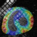
Quantitative analysis and modeling of myocardial function in MRI
Cardio-vascular diseases are the leading cause of mortality in industrialized countries, and therefore a major public health challenge. As a result of intensive technological developments, Magnetic Resonance Imaging (MRI) is today recognized as a relevant modality for dynamically imaging the heart anatomy and function, and provides a valuable investigation tool for early diagnosis and clinical / therapeutical follow-up. The CardioMeter project aims at developing and validating advanced image analysis methods for the automated measurement of myocardial and wall motion from MRI sequences. It also aims at elaborating patient-specific, comprehensive compact models of the myocardial contracile function for the non-pathological heart as well as for specific pathologies.
ARTEMIS contributions concern: (1) the development of a Computer-Assisted Diagnosis (CAD) tool for the quantitative analysis of tagged MRI exams, automatically delivering deformation parameters at the voxel, myocardial segment and whole myocardium scales; and (2) the generation of statistical motion atlases allowing for a compact parametric modelling of myocardial deformations. Based on novel variational statistical non rigid registration techniques, these results have been clinically validated for ischemic and hypertrophic cardiomyopathies.
![]() Partners: APHP - Hôpital Pitié-Salpêtrière / Service de Radiologie
Partners: APHP - Hôpital Pitié-Salpêtrière / Service de Radiologie
![]() Keywords: Computer-Aided Diagnosis, Medical imaging, Modeling, Registration, Segmentation
Keywords: Computer-Aided Diagnosis, Medical imaging, Modeling, Registration, Segmentation

![[del.icio.us]](https://artemis.telecom-sudparis.eu/wp-content/plugins/bookmarkify/delicious.png)
![[Digg]](https://artemis.telecom-sudparis.eu/wp-content/plugins/bookmarkify/digg.png)
![[Facebook]](https://artemis.telecom-sudparis.eu/wp-content/plugins/bookmarkify/facebook.png)
![[Google]](https://artemis.telecom-sudparis.eu/wp-content/plugins/bookmarkify/google.png)
![[Jamespot]](https://artemis.telecom-sudparis.eu/wp-content/plugins/bookmarkify/jamespot.png)
![[LinkedIn]](https://artemis.telecom-sudparis.eu/wp-content/plugins/bookmarkify/linkedin.png)
![[MySpace]](https://artemis.telecom-sudparis.eu/wp-content/plugins/bookmarkify/myspace.png)
![[Reddit]](https://artemis.telecom-sudparis.eu/wp-content/plugins/bookmarkify/reddit.png)
![[Technorati]](https://artemis.telecom-sudparis.eu/wp-content/plugins/bookmarkify/technorati.png)
![[Twitter]](https://artemis.telecom-sudparis.eu/wp-content/plugins/bookmarkify/twitter.png)
![[Email]](https://artemis.telecom-sudparis.eu/wp-content/plugins/bookmarkify/email.png)